
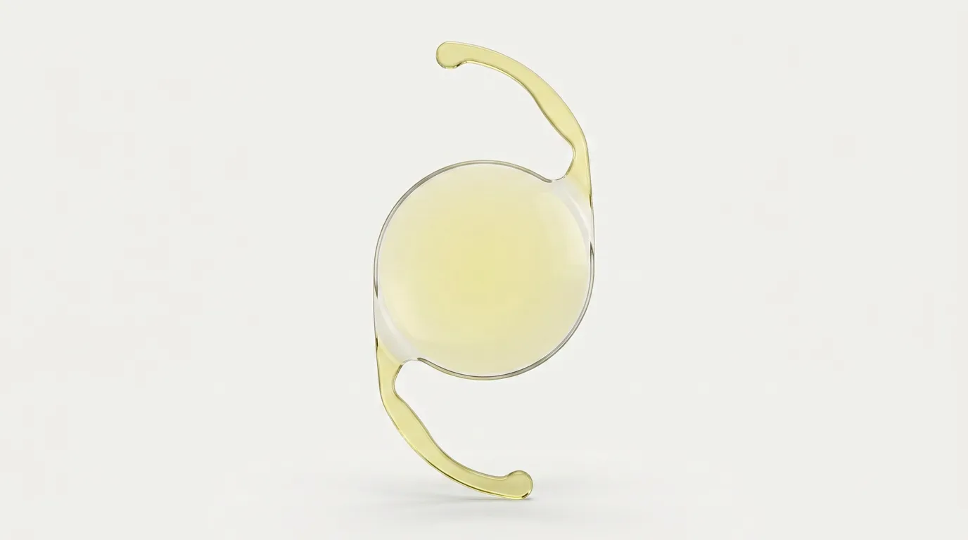
Once a patient has accepted that cataract surgery lies ahead, the same question almost always follows. It is not, "Will it hurt?" and it is not, "How long does it take?" It is: "Doctor, I have heard that there are better lenses that mean I will not need glasses. Is it worth paying extra for one?"
This conversation takes place at every preoperative cataract assessment at VIDAR-ORASIS SWISS Eye Hospital in Novi Sad. The clinic has existed since 2010 and its team includes four university professors. What follows covers the same subjects discussed during that conversation, including all the compromises a patient should understand before making a decision.
The question is entirely legitimate and deserves an honest answer rather than a sales answer. Unfortunately, the answer is longer than one sentence because there is no single lens that is best for everyone. There is a lens that is most appropriate for your eye, your way of life and your habits, and those three things differ from one person to another. Someone who works at a computer all day and drives at night has completely different needs from someone who reads for hours in an armchair and almost never drives after dark.
This article explains the differences between artificial lens types in the way they would be explained in the consulting room, including all the compromises. We will cover monofocal lenses, multifocal lenses, extended-depth-of-focus lenses, aspheric designs and lenses that correct astigmatism. You will see a table showing what can realistically be expected without glasses with each lens type. You will also learn about halos, which are rarely mentioned in sales materials, and about eye characteristics that make certain lenses an unreasonable option.
One point should be placed in its proper context immediately. The purpose of cataract surgery is not to free you from glasses. Its purpose is to remove the cloudy lens that no longer transmits light properly and replace it with a clear lens of appropriate power. Independence from glasses is an additional possibility, not a promise. We have explained when surgery is indicated in our detailed guide to the timing of cataract surgery, while basic information about the procedure is available on our Cataract Surgery page.
What follows is not a recommendation for your individual case. A specific lens can be selected only after an examination and measurements because the choice depends on findings that cannot be seen without specialised instruments.
What an artificial lens can and cannot do
Before comparing lens types, it is important to understand what actually happens inside the eye during surgery. Without that, a comparison table has little meaning.
Why is the lens replaced?
The natural lens sits inside the eye immediately behind the pupil and is approximately the size of a bean. In a child, it is completely transparent and highly flexible. Together with the cornea, the transparent front surface of the eye that can be compared to the glass on a watch, it refracts light and focuses an image onto the retina. The retina is a thin layer of nervous tissue at the back of the eye and performs a role similar to a camera sensor.
A cataract is a clouding of this natural lens. It cannot be made clear again with eye drops, exercises or diet. The only way to restore transparency is to remove the cloudy lens and replace it with an artificial lens, known in medical terminology as an intraocular lens or IOL. You can read more about the condition itself on our Cataract page.
What an artificial lens does not do
This is the most important part of the entire article and the source of the greatest number of misunderstandings.
The natural lens in a young eye changes shape. When you look into the distance, it is stretched and flatter. When you look at a book, the small muscle surrounding it contracts, the lens becomes rounder and refracts light more strongly, moving the focus to near vision. This ability is called accommodation and is the reason a child can read text ten centimetres from the nose.
Accommodation gradually weakens in everyone and usually becomes noticeably limiting around the age of 40. That is when reading glasses become necessary. The change is called presbyopia, and we have discussed it separately in our guide to reading glasses after the age of 40.
No artificial lens routinely implanted today changes shape inside the eye in the way the natural lens did. Its optical system is fixed. This means that focusing at different distances has to be achieved through an optical strategy built into the lens design rather than true focusing. Every one of these strategies has a cost, and the remainder of this article explains those costs.
What else determines the final result?
An artificial lens is an optical device. It may focus an image perfectly, but if the structure receiving that image is damaged, the result will not be the same as it would be in a healthy eye.
Before surgery, the doctor therefore examines more than the lens. The retina is assessed, particularly its central part, the macula, which is responsible for sharp central vision. The optic nerve is examined because glaucoma damage causes permanent loss of the visual field. The cornea is assessed for transparency, shape and the number of cells on its inner surface. The tear film is also considered because dry eye can reduce image quality before and after surgery.
A problem in one of these structures does not necessarily mean that surgery cannot be performed. More often, it means that expectations must be adjusted and the lens choice becomes narrower because certain lens types require a healthy retina to provide any meaningful benefit.
Biometry: the measurement that determines lens power
One common misunderstanding should be resolved immediately. Lens type and lens power are two different decisions.
Lens type answers the question of how the lens distributes light, including whether it has one focus, several focuses or an extended range of focus. Lens power, expressed in dioptres, answers how strongly the lens refracts light and therefore at which distance the image will be sharp.
You choose the lens type together with the surgeon according to your way of life. Nobody simply chooses the power. It is calculated from measurements of your eye, and that process is called biometry. You can read more about the examination on our Ocular Biometry page.
What is measured?
Several measurements of your eye are needed to calculate the lens power.
The first is the axial length of the eye, the distance from the front surface of the cornea to the retina. This is the single most important measurement. Human eyes are not all the same size. They are generally longer in short-sighted people and shorter in long-sighted people, and a difference of one millimetre changes the required lens power by several dioptres. It is therefore measured with a level of precision not achieved by an ordinary eye examination.
The second measurement is corneal curvature, often recorded as K1 and K2, which are values for the two principal meridians. The cornea is the most powerful optical surface of the eye. If it is evenly rounded like half a ball, it refracts light equally in all meridians. If it is more curved in one meridian, like the back of a spoon, astigmatism is present. We discuss this in a separate section.
These measurements are combined in a mathematical formula to calculate the lens power required to achieve the agreed visual target.
Optical and ultrasound biometry
Biometry can be performed in two ways.
Optical biometry uses a beam of light. The patient sits in front of the instrument, rests the chin, looks at a target and the measurement takes only a short time. The eye is not touched. This is the first-choice method for most patients because it is accurate, repeatable and comfortable.
Ultrasound biometry uses a sound wave. After a local anaesthetic drop has been applied, a probe is placed against the eye, or a technique is used in which fluid remains between the probe and the eye. This method is used when the cataract is so dense that the light beam cannot pass through it to the retina or when the patient cannot fix their gaze for another reason.
The method used in your case depends on the condition of the eye and the available equipment. You will receive an answer in the consulting room. The measurement itself is not painful, requires no preparation at home and has no lasting effects.
Why is no calculation absolutely precise?
This is something patients rarely hear in advance but should understand.
The formula predicts where the lens will sit inside the eye from the measured parameters. Its actual position after surgery also depends on how the capsule, the thin membrane in which the lens is placed, behaves during the weeks of healing. The capsule contracts slightly and may move the lens by a fraction of a millimetre forwards or backwards. This movement cannot be measured in advance in any patient.
As a result, there can be a difference between the planned and achieved prescription. In most patients, this difference is small and is barely noticed. Some patients retain a small prescription, commonly up to approximately one dioptre. This may be corrected with light distance glasses or, if it causes no difficulty, simply left uncorrected.
This is not an error and it is not a complication. It is a limit of the method's precision and should be discussed before surgery rather than afterwards. Be cautious when someone promises an exact prescription to a decimal place because that degree of precision is not achievable in biology.
A small remaining prescription usually causes little difficulty with a single-focus lens. The same prescription may be more noticeable with a multi-focus lens because it disrupts the fine distribution of focus on which the design depends. This is one reason multifocal lenses are selected more cautiously.
If you have previously had laser vision correction
Calculation is substantially more complex in one group of patients: people who have previously had PRK or LASIK laser vision correction.
The reason becomes logical once explained. Standard formulas assume a particular relationship between the front and back surfaces of a cornea that has not been treated. Laser correction changes the shape of the front surface while leaving the back surface unchanged. That assumed relationship no longer applies, so using the formula without adjustment produces an incorrect lens power.
Old medical records are therefore extremely useful in these patients, including the prescription before laser correction, corneal topography and details of the procedure. If you have them in a drawer, bring them. If they are unavailable, methods exist that can calculate without them, but every additional piece of information helps.
Monofocal lens
A monofocal lens has a single focus. It is the optically simplest lens and directs the light passing through it into one focal plane.
What does one focus mean in practice?
Because the lens has one focus, its location must be chosen. This decision is made with you before surgery.
The most common choice is distance focus. After recovery, this generally means watching television, recognising people in the street, driving and moving around without glasses. Reading, telephone use, fine work and anything closer than approximately arm's length require near-vision glasses. These glasses are usually simple and readily available. Less commonly, people who have been short-sighted throughout life and are accustomed to reading without glasses deliberately choose near focus. In both cases, the same type of lens is used. Only the calculated power differs.
Who is a monofocal lens most suitable for?
A monofocal lens is a reasonable choice for the great majority of patients and should not be viewed as an inferior option. It provides the cleanest image for the available amount of light because it does not divide light between focuses, and it has the smallest effect on contrast sensitivity.
It is particularly suitable for people who drive frequently at night, people who work in low-light conditions, people with any changes affecting the retina or optic nerve, and everyone who is comfortable using reading glasses. If glasses are not an inconvenience but a habit of the past 30 years, a monofocal lens is unlikely to disappoint.
Monovision: one eye for distance and the other for near vision
There is an approach used with monofocal lenses that deserves an explanation because it is mentioned frequently.
The idea is to set one eye, usually the dominant eye, for distance and the other for near vision or mild short-sightedness. The brain then uses the eye that provides the clearer image for each situation and suppresses the other image. When the difference between the eyes is mild, this is described as soft or partial monovision.
The advantage is reduced dependence on glasses while retaining simple optics in both eyes. The disadvantage is that some people feel that depth perception is different, while a smaller number simply cannot tolerate the difference between the eyes. Whenever possible, the effect is therefore tested first with contact lenses.
This is not a solution for everyone and is not chosen lightly, but it can be an elegant option in the right patient.
Multifocal lens
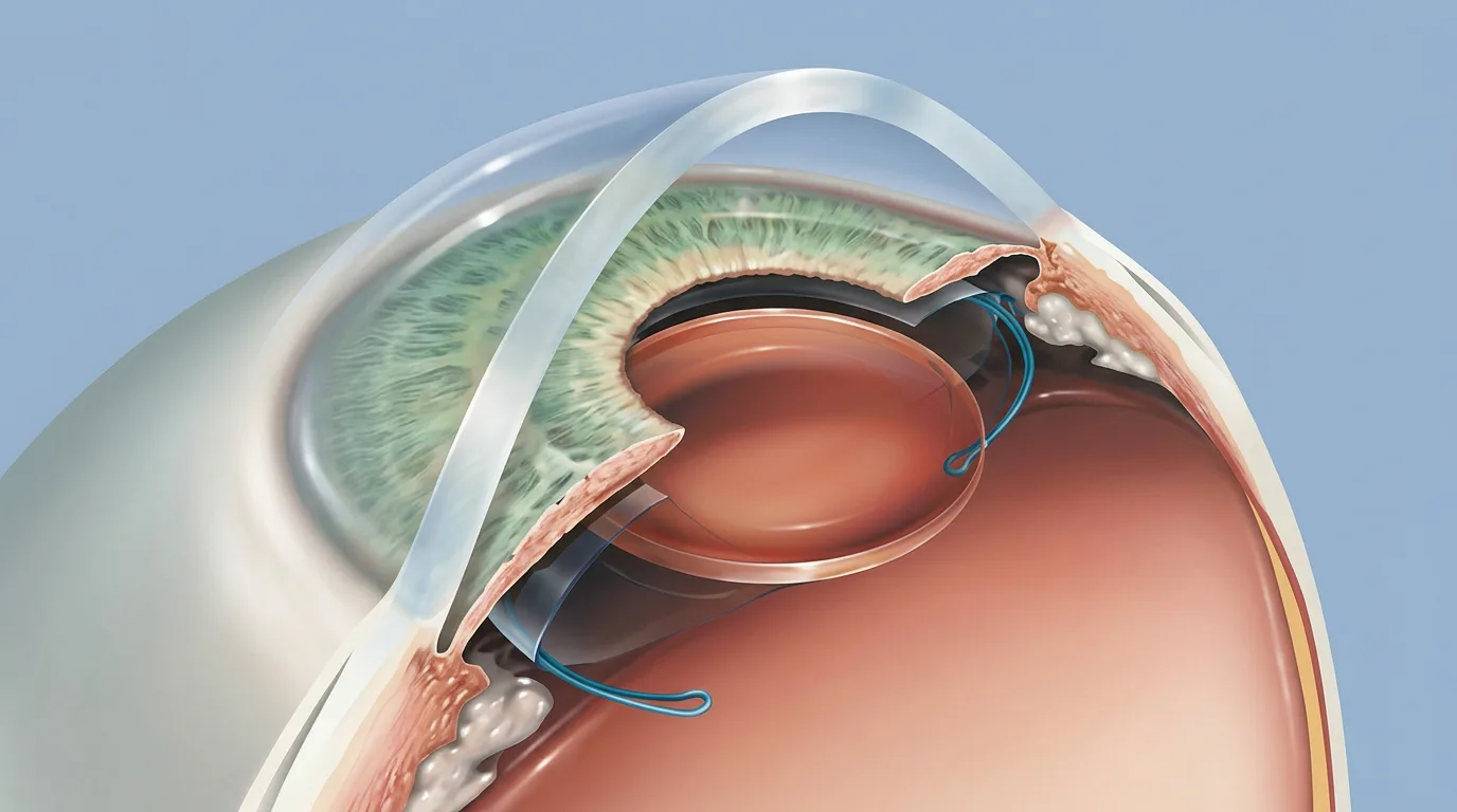
A multifocal lens has several focal points. It is designed to respond to the wish to use glasses as little as possible after surgery.
How light is divided
The optical surface of a multifocal lens resembles a series of concentric rings. These rings divide incoming light into several beams and direct them into different focal planes. Part of the light creates a sharp image for distance, part for near vision and, in newer designs, part for intermediate vision.
At every moment, one sharp image and one or more blurred images therefore exist simultaneously inside the eye. Over time, the brain learns to pay attention to the image that is useful in that situation and suppress the others. This is a genuine process and takes time, as we explain below.
The advantage and disadvantage follow directly from this description. The advantage is useful vision at several distances without glasses. The disadvantage is that none of the images receives all the available light and the blurred images are not completely invisible.
What patients describe as the benefit
When the choice has been appropriate and recovery is uncomplicated, patients with multifocal lenses describe specific everyday benefits.
They get up in the morning and see the clock on the wall without looking for glasses on the bedside table. They read a message on the telephone while walking. In a shop, they turn a package over and read the expiry date without taking glasses from a bag. At work, they move their gaze from the monitor to the person opposite and back without repeatedly removing and replacing glasses. They play with a grandchild, read a picture book and then look out onto the balcony.
This is a realistic picture of what can be gained: not perfection, but reduced dependence on an object that is constantly searched for and misplaced. For many people, that is valuable, and it is fair to say so.
Halos, glare and contrast
We now come to the part that is rarely discussed in advance but must form part of the conversation.
A halo is a ring of light around a bright light source in the dark. The classic description involves night driving: headlights from oncoming vehicles, streetlights and traffic signals acquire a ring around them. The cause is the blurred image that always exists alongside the sharp image in multifocal optics. Around a point source of light in darkness, it appears as a circle.
Glare is the scattering of light that reduces clarity around a bright source. It occurs with opposing light, on a wet road at night and sometimes in strong sunlight.
Reduced contrast sensitivity is the third and most subtle effect. Contrast sensitivity is the ability to distinguish an object from its background when the two are not black and white but similar in brightness: a grey stair against a grey floor, the edge of a pavement on a cloudy day, or a face in dim light. Because multifocal optics divide light, contrast sensitivity is generally somewhat lower than with simple single-focus optics. This may not be visible on a letter chart because the chart is black and white and brightly illuminated. It may occasionally be noticed in daily life.
In most patients, these effects are mild and become less noticeable over time. In some patients, they remain noticeable. In a small number, they cause sufficient difficulty for replacement of the lens to be considered. That is another operation with its own risks.
The question before choosing a multifocal lens is therefore not only, "Would you like to be free from glasses?" It is also, "How would you feel if you saw rings around headlights at night?"
The period of adaptation
The brain has to learn to work with the new optical system. This process is called neuroadaptation.
During the first days and weeks after surgery, many patients describe the image as unusual, report that they need to find the correct reading distance, and notice halos. In most people, this gradually settles over several weeks to several months.
Two things help. The first is knowing in advance that this may happen because an expected effect is far less alarming. The second is patience, because judging the result after ten days is not meaningful. We have explained the postoperative period separately in our guide to recovery after cataract surgery.
Discuss the lens choice with the surgeon. Call 021 63 61 222 to book an examination at VIDAR-ORASIS SWISS Eye Hospital, Bulevar oslobodjenja 76A, Novi Sad. We are open on weekdays until 20:00. The examination includes ocular biometry, a discussion of your daily routine and an explanation of which options make sense for you and which do not.
Extended-depth-of-focus lens (EDOF)
Between a single-focus lens and a lens with several separate focal points lies a third approach that has become increasingly common in ophthalmology. These are extended-depth-of-focus lenses, described in the international literature by the abbreviation EDOF.
How does it differ from a multifocal lens?
A multifocal lens creates two or three separate points of sharp vision, with a less distinct area between them. Imagine two lights switched on in a corridor with darkness between them.
An extended-depth-of-focus lens creates one elongated range in which the image remains usefully sharp instead of two separate points. Rather than two lamps, imagine one longer strip of light. There is no pronounced gap between distance and intermediate vision, so the transition feels smoother and more natural.
The price of this solution is that the range does not extend indefinitely. In most designs, good sharpness covers distance and intermediate vision, including driving, moving around, watching television, using a computer, looking at a board on the wall and reading prices on a shelf. Small print at near distance often still requires glasses. A small product label, telephone directory, medicine instructions and a book with small print commonly remain tasks for which help is needed.
Who is this approach usually suitable for?
The reasoning is clear when described in practical terms. If most of your day takes place between half a metre and infinity, including computer work, meetings, driving, cooking and moving around indoors and outdoors, while reading small print is occasional, that pattern matches extended focus well.
If reading is your main daily activity and small print is central to your work, this lens is less likely to meet expectations because you may still reach for glasses in the situation where they bother you most.
Compromises you should understand
Halos and glare are generally less pronounced with extended-depth-of-focus lenses than with traditional multifocal lenses, but they are not excluded. Some patients describe a different effect, with light spreading in a subtle star pattern instead of a regular ring.
Contrast sensitivity is usually better preserved than with multifocal lenses and less well preserved than with simple single-focus optics.
The rule about remaining prescription also applies. Extended focus tolerates a small difference from the planned prescription to some extent, but not without limit, so measurements are performed just as carefully.
Important: this description concerns a type of lens used in modern ophthalmology in general. Whether a specific EDOF lens is implanted at VIDAR-ORASIS SWISS is something you need to confirm in the consulting room. Do not plan your decision from this article before checking by telephone or during an examination.
Aspheric lens
Asphericity does not refer to the number of focuses. It is a property of the lens surface. It is therefore not chosen instead of a monofocal or multifocal lens, but may be a feature of either.
What does an aspheric surface mean?
A traditional spherical surface is part of a ball and has the same curvature at every point. Such a surface has an inherent limitation: rays passing through its edge focus slightly differently from rays passing through the centre. This is called spherical aberration and creates a small amount of image spread, most noticeable when the pupil is wide in darkness.
An aspheric surface is not part of a ball. Its curvature changes slightly from the centre towards the edge to reduce that error.
What does the patient notice?
The effect is most noticeable in low light when the pupil is wider and more light passes through the edge of the lens. Patients may describe a somewhat cleaner image at night and less spreading around lights.
Toric lens for astigmatism
To explain the purpose of this lens, we first need to explain astigmatism because the term is frequently misunderstood.
What is astigmatism?
A regular cornea is rounded equally in all meridians like half a football. With astigmatism, it is more curved in one meridian than another, more like the back of a spoon or a rugby ball.
As a result, the eye does not have one point of focus but two in different directions. The image is stretched. Patients may describe elongated letters, a shadow or double outline around bright objects, or the need to squint to improve focus. Almost every eye has a small amount of astigmatism, which usually causes no difficulty. Once it exceeds a certain level, it reduces image quality regardless of how accurately the rest of the optical system is adjusted.
This matters in cataract surgery for one reason. Replacing the lens corrects clouding and refractive power, but it does not change the shape of the cornea. If the astigmatism comes from the cornea, it remains after surgery.
How is astigmatism managed during cataract surgery?
Two approaches are commonly used in ophthalmology.
The first and simplest is to leave the astigmatism uncorrected during surgery and use glasses with a cylindrical correction afterwards. This is entirely acceptable for many patients, particularly if they plan to use reading glasses anyway.
The second approach is to implant a lens with toric optics. Such a lens is deliberately designed to refract light more strongly in one meridian than another, by exactly the amount needed to neutralise the corneal irregularity. To work, it must be positioned at a precise axis inside the eye and remain at that axis. If it rotates over time, part of the effect is lost and an additional procedure to return it to the correct position may sometimes be considered.
The appropriate approach depends on the amount of astigmatism, whether it is regular or irregular, and the findings of corneal topography, a measurement that precisely maps the corneal shape.
Why is irregular astigmatism a special case?
Regular astigmatism has two principal meridians at right angles to each other and can be corrected optically with glasses or a toric lens.
Irregular astigmatism, for example after corneal scarring, in keratoconus or following certain previous procedures, cannot be fully neutralised by glasses or an implanted lens because its irregularity cannot be described by simple geometry. These eyes require individual planning and different expectations.
Important: this section also describes what is used in ophthalmology in general. Confirm during the examination or by telephone whether toric lenses are implanted at VIDAR-ORASIS SWISS.
Table: what you can expect to see without glasses
The following table summarises what has been explained so far. Before reading it, there are three qualifications that are not merely formal disclaimers.
First, the descriptions are indicative and represent typical behaviour of groups of lenses, not the result in your eye. Differences between individual designs within the same group may sometimes be greater than the table suggests.
Second, every description assumes a healthy retina, a healthy optic nerve and uncomplicated recovery. In an eye with another condition, the result is determined by that condition as well as the lens choice.
Third, the price column contains no figures because additional charges vary according to lens type and supplier. Current information is available only from the clinic or by telephone. The price of standard cataract surgery appears on the Services and Price List page. Any additional charge for a lens with extra optical properties is not stated in this article and must be confirmed with the clinic.
| Lens type | Distance | Intermediate vision | Near vision | Need for glasses | Night vision and halos | Additional charge |
|---|---|---|---|---|---|---|
| Monofocal, set for distance | very good | limited | poor | reading glasses almost always required | least pronounced halos, best preservation of contrast | no additional charge, included in standard surgery |
| Monofocal, set for near vision | poor | limited | very good | glasses required for distance and driving | same as above | no additional charge |
| Monofocal with monovision | good in the dominant eye | often useful | good in the other eye | occasionally required for prolonged reading and night driving | halos are not pronounced, but depth perception may feel different | usually no additional charge, confirm with the clinic |
| Aspheric design, a property rather than a separate lens type | depends on the lens type | depends on the lens type | depends on the lens type | depends on the lens type | a somewhat cleaner image in darkness | depends on the model, confirm with the clinic |
| Extended depth of focus (EDOF) | very good | very good | limited, small print usually requires glasses | glasses mainly required for small print | halos are milder than with multifocal lenses but remain possible | additional charge, confirm the amount with the clinic |
| Multifocal | good | good with newer designs | good | glasses required occasionally or rarely | most pronounced halos and glare, with somewhat lower contrast | additional charge, confirm the amount with the clinic |
| Toric, for astigmatism and combined with one of the lens types above | depends on the underlying lens type | depends on the underlying lens type | depends on the underlying lens type | reduces the need for a cylindrical correction in glasses | depends on the underlying lens type | additional charge, confirm the amount with the clinic |
The table covers lens types used in modern ophthalmology in general and is not a list of what is available at VIDAR-ORASIS SWISS. Confirm which types the clinic implants by telephone or at the examination.
Read the table as a map of compromises, not a ranking. The first row is not the worst option, and the last row is not the best. Every row represents a good choice for someone.
Who is not a good candidate for a multifocal lens?
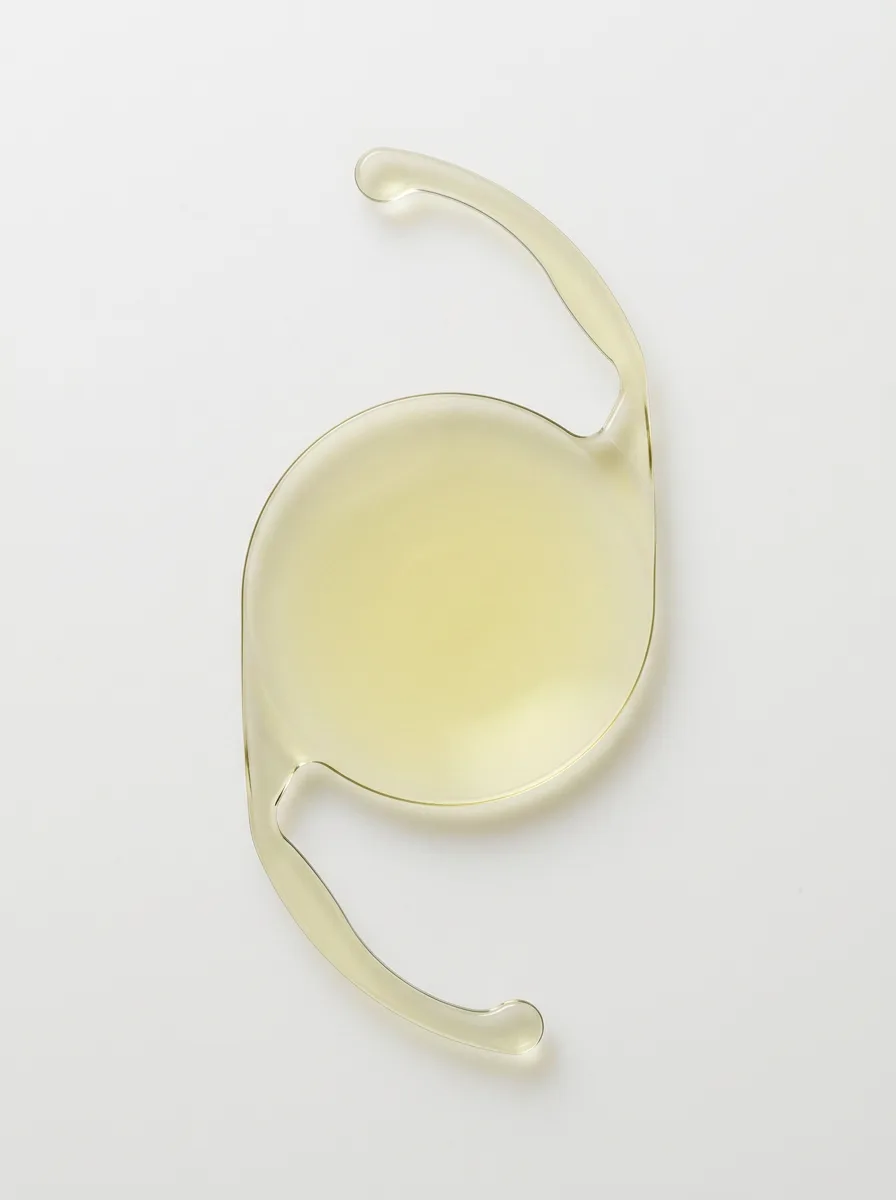
There are conditions in which multi-focus lenses are generally not recommended because the disadvantage may outweigh the benefit. This is not a list of absolute prohibitions. It is a list of reasons for caution, and the final assessment is made by the doctor after examination.
Diseases of the macula
The macula is the central part of the retina responsible for sharp vision. In macular degeneration, macular swelling from any cause and other changes affecting this area, the quality of central vision is already reduced.
Multifocal optics divide light and further reduce contrast. In an eye that already has a macular problem, this means using some of the remaining image quality for a feature that may not provide a benefit. A single-focus lens, which directs all available light into one image, is therefore generally chosen for these eyes.
Glaucoma with visual field damage
Glaucoma damages the optic nerve and narrows the visual field, often without being noticed for years. In a patient who already has visual field loss, an additional reduction in contrast sensitivity can make orientation more difficult, especially in dim light and on stairs.
Irregular astigmatism and corneal disease
Multifocal optics work as intended only when light reaches the lens through a regular cornea. With irregular astigmatism, scarring, keratoconus or reduced corneal transparency, the image is already distorted before it reaches the lens, so the finely arranged focal points lose their purpose.
Professional drivers, especially those who drive at night
The decision is made very cautiously in this group. If the quality of night vision affects your work and the safety of other people, halos and glare are not an aesthetic inconvenience but a serious practical issue.
Unrealistic expectations
This is the most sensitive point and must be said aloud. A patient who arrives saying, "I never want to wear glasses again and I want vision like I had at 20," is not a good candidate for any lens until those expectations have been aligned with reality.
A multi-focus lens reduces the need for glasses. It does not reliably eliminate glasses for everyone and it does not return the eye to youth. People who accept this are generally satisfied. Those who do not may remain dissatisfied even after technically perfect surgery.
How we choose together with you
Lens selection is not a moment in which the doctor simply states a number. It is a structured conversation.
First, we assess what the eye allows
Before discussing preferences, the examination answers what is medically and optically possible. The retina and macula, optic nerve, cornea, eye pressure, pupil size and reaction, and tear film are assessed. Biometry is performed.
This produces a list of options that are reasonable for your eye. In some patients, the list is long. In others, it contains only one option. Both outcomes are normal.
We then consider what your day looks like
It is useful to describe an ordinary day to your doctor from morning to evening. How many hours do you spend at a screen? Do you drive, how often and at what time of day? Do you read books or mainly use a telephone? Do you perform close manual activities that require sharp near vision, such as embroidery, fishing, fine repairs or playing cards? Do glasses create practical problems, for example by fogging when you come in from the cold?
We then choose the compromise and record the agreement
When the possibilities allowed by the eye are considered alongside your habits, one or two reasonable solutions usually remain. The refractive target is then agreed: whether the aim is clear distance vision with reading glasses or another combination.
Good practice is to repeat aloud at the end of the conversation what has been agreed and what can realistically be expected, including the possibility of a small remaining prescription. If you leave without a clear understanding of what you are likely to do without glasses, return to the question. That is not being difficult. It is sensible preparation.
If both eyes are operated on
Cataract develops in both eyes in most patients, so the operations are performed separately with an interval between them. The experience from the first eye is valuable because it is only then that you know how the optical system feels in real life.
When there is no other reason to hurry, the decision for the second eye may therefore be made after the first eye has recovered. If the first eye works well with the selected lens type, the same approach is generally used for the second.
Price, additional charges and what the package includes
Patients often leave this question until the end of the conversation even though it belongs near the beginning.
The price of standard cataract surgery is stated on the Services and Price List page. Prices are subject to change and should always be confirmed by telephone before booking. Biometry is charged as a separate measurement and is also listed in the price list.
An additional charge for a lens with extra optical properties, if such a lens is chosen, is not included in the standard surgery price. The amount depends on the lens type and is available only from the clinic. This article deliberately gives no estimate for additional charges because any such figure would be unverified.
Discuss the lens choice with the surgeon. Call 021 63 61 222 to book an examination at VIDAR-ORASIS SWISS Eye Hospital, Bulevar oslobodjenja 76A, Novi Sad. We are open on weekdays until 20:00. Bring a list of the situations in which vision matters most to you and any previous eye records, particularly if you have had laser vision correction.
What to ask your doctor
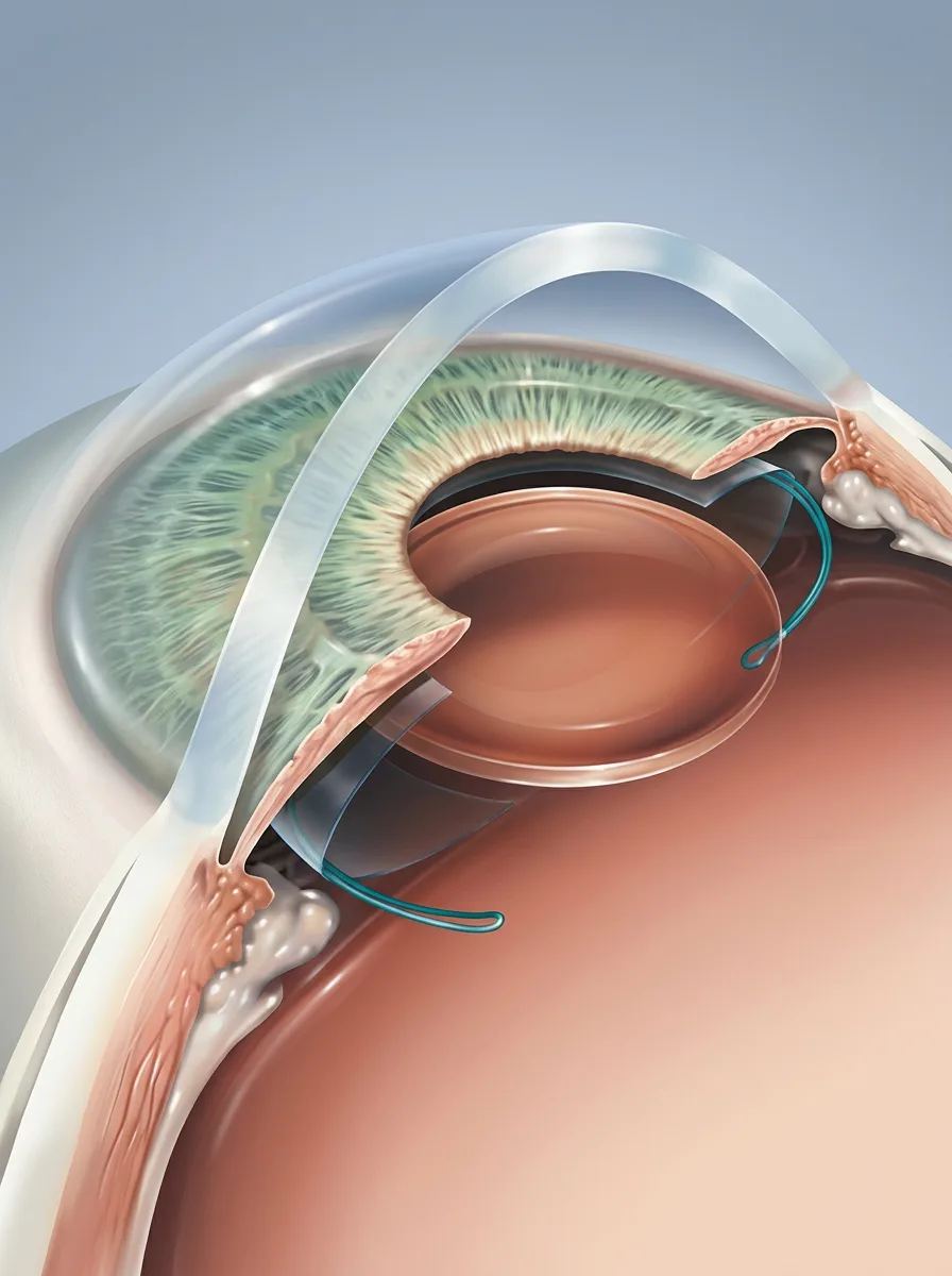
The following questions are useful regardless of which lens you ultimately choose. Write them down and bring them with you.
Does the condition of my retina and optic nerve allow me to consider a multi-focus lens?
Which lens types does the clinic implant, and which of them are realistic options for me?
At which distance are we planning the focus, and what does that specifically mean for reading and driving?
How likely is it that a small prescription will remain after surgery, and what happens if it does?
If halos cause serious difficulty, what options would be available?
What is included in the price, how much is any additional charge, and when is it paid?
Frequently asked questions
Is it worth paying extra for a lens that may reduce my need for glasses?
It depends on how much glasses genuinely interfere with your life and how you feel about the compromises such lenses bring. If reading glasses do not trouble you, paying extra may not solve a problem you actually have. If independence from glasses is important, your retina is healthy and you do not drive frequently at night, it is reasonable to discuss the option. The decision should not be made before an examination.
Does a multifocal lens guarantee that I will not need glasses?
No. That cannot be promised. Some patients no longer need glasses in almost all situations, while others still use them for small print or prolonged reading. The realistic goal is a reduced need for glasses, not a guaranteed elimination of them.
What exactly are halos, and how long do they last?
They are rings of light around bright sources in darkness, most commonly headlights and streetlights. They occur because multifocal optics simultaneously create a blurred image alongside the sharp one. In most patients, they become less noticeable over several weeks to several months, but in some they remain.
Can the lens be replaced if it does not suit me?
Replacement is possible, but it is another operation with its own risks and is not performed routinely. This is why the conversation before the first operation matters so much. Before considering replacement, sufficient time is usually allowed for neuroadaptation and other causes are checked, such as dry eye or clouding of the capsule behind the lens.
Does an artificial lens become cloudy over time?
The implanted lens does not become cloudy. In some patients, the thin capsule holding the lens becomes cloudy over time, causing symptoms similar to cataract. This is called posterior capsule opacification or secondary cataract and is treated with a short laser procedure. We have explained it in our separate guide to secondary cataract.
Our article on the timing of cataract surgery explains when surgery is considered and how the appropriate time is assessed. Our recovery guide explains the period after surgery.
I previously had laser vision correction. Does that change anything?
It changes two things. Calculating lens power is more complex, and the risk of a remaining prescription is greater, so old records are very useful. In addition, the altered corneal shape may narrow the range of suitable lens types in some patients. This does not mean surgery is impossible. It means preparation requires more information.
How long after surgery should I wait before getting new glasses?
The prescription settles only as the eye heals, so glasses are not usually prescribed immediately. The exact time is determined by the doctor during follow-up, commonly at one of the examinations in the first weeks after surgery.
Important note
This article is for information only and does not replace an examination by an ophthalmologist. The descriptions concern lens types used in modern ophthalmology in general and do not confirm that every listed type is available at VIDAR-ORASIS SWISS. A lens cannot be selected from an article. It can be chosen only after an examination and measurements. Confirm prices and any additional charges by telephone because they are subject to change.
Sources and further reading
The medical content was reviewed by Prof. Dr Mirko Resan. This information does not replace an eye examination or individual advice from an ophthalmologist.