
Eye clinic / patient information
Ocular ultrasound examination
Ocular ultrasound is used when the fundus cannot be seen clearly. B-scan assesses retinal detachment and vitreous haemorrhage, while A-scan measures the eye before cataract surgery.
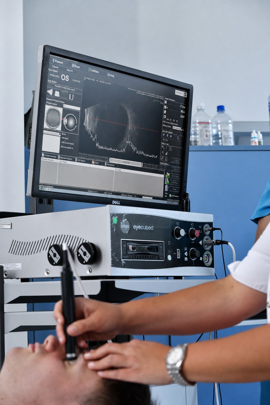
The eyeball and the orbit are anatomically well suited to the use of ultrasound (US) for diagnostic purposes. Ultrasound waves are mechanical vibrations (above 16 KHz) that are transmitted well through solid and liquid media. The probe of the ultrasound device emits and receives the reflected ultrasound vibrations. There are two methods of using ultrasound in ophthalmology:
1. A scan. This is a one dimensional method for recording an echogram (an ultrasound trace) in the form of a line. It is most often used for biometry in the calculation of the intraocular lens that is to be implanted during cataract surgery.
2. B scan. This is a two dimensional method, and this echogram consists of many points of differing density and brightness. It is used for the topographic diagnosis of intraocular changes and orbital processes. The B scan is important in the diagnosis of: retinal detachment, retinoschisis, bleeding into the vitreous cavity, proliferative diabetic retinopathy, intrabulbar and intraorbital tumours, intrabulbar foreign bodies, thickening of the extraocular muscles in thyroid eye disease, and similar conditions.
Appointments
Not sure which examination you need?
Tell us about your symptoms and our team will help you book the right first step.