
Eye clinic / patient information
Surgical treatment of keratoconus
Keratoconus is a disease of the cornea in which the corneal tissue loses its rigidity, changes into a cone shape and becomes thinner, which leads to irregular astigmatism and a consequent decline in vision.
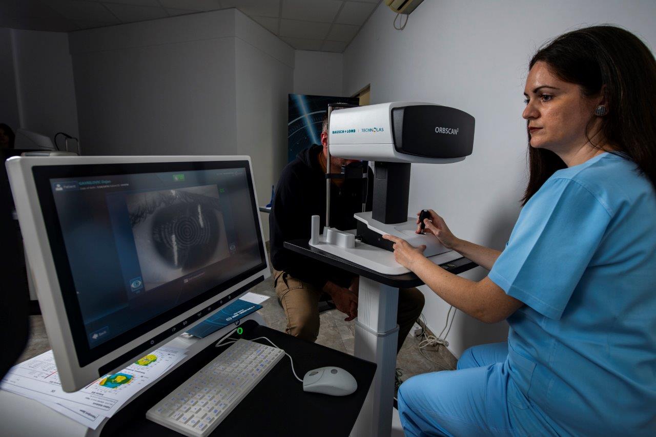
If keratoconus progresses and threatens a greater thinning of the cornea, then it is necessary to perform one of the surgical methods by which the cornea will be strengthened (“cemented”) and its further thinning prevented.

- Corneal cross-linking (CXL) is a surgical method that we use when the disease is in the phase of progression and when there is still a certain thickness of tissue for this procedure. Through the action of a vitamin B2 solution and special UV lamps, we create “cross-links” in the basic network of collagen fibres, and thereby we strengthen the biomechanical resistance of the cornea.
- When the cornea is altered by scarring, we then proceed to the operative procedure which is called keratoplasty. We remove the diseased tissue of the cornea and replace it with donor tissue of full thickness (penetrating keratoplasty). Recently, the approach of lamellar keratoplasty (DALK) has also been used in specially selected cases.
Appointments
Not sure which examination you need?
Tell us about your symptoms and our team will help you book the right first step.