
Eye clinic / patient information
Specular microscopy
Specular microscopy assesses the corneal endothelium before cataract surgery and supports the diagnosis and monitoring of corneal disease.
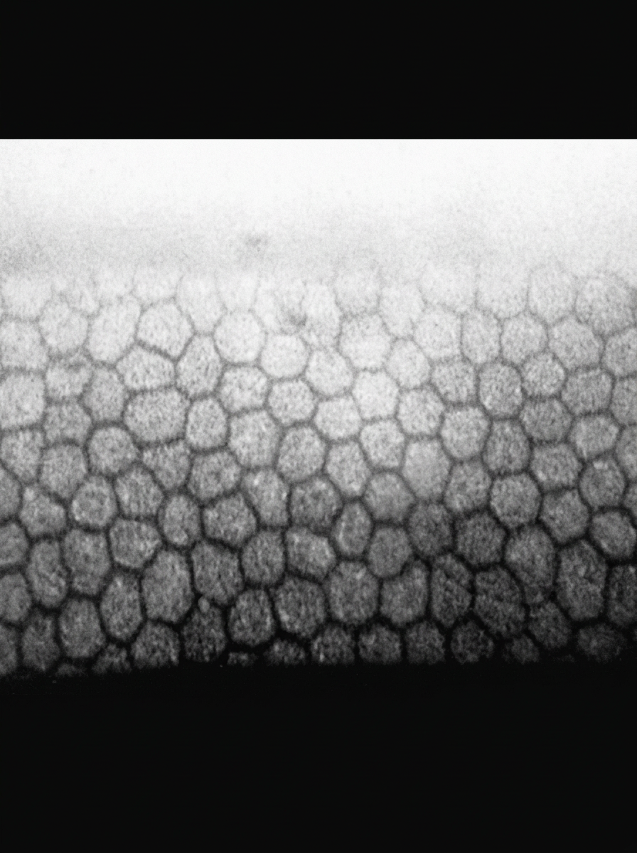
Specular microscopy is a non-invasive, non-contact method that is essential in diagnosing conditions affecting the corneal endothelium. The corneal endothelium is a vital layer that enables the cornea to function normally and remain transparent. It is located on the back surface of the cornea and consists of a single layer of metabolically active cells. These cells help maintain the normal amount of water in the cornea and, consequently, its normal transparency.
Specular microscopy is used to analyse the corneal endothelium and calculate the exact number, appearance and size of endothelial cells. If an examination shows a reduced number of these cells or irregularities in their structure, such as age-related changes known as cornea guttata, this method is particularly important.
Before cataract surgery, especially when the cataract is hard and brunescent and cornea guttata is also present, it is advisable to determine the exact endothelial cell count. This is important when assessing how the cornea may respond after cataract surgery, during which ultrasound energy is used.
Specular microscopy is also important in diagnosing and monitoring Fuchs endothelial corneal dystrophy, as well as in the assessment and postoperative follow-up of corneal transplant surgery, including penetrating or lamellar keratoplasty.
Appointments
Not sure which examination you need?
Tell us about your symptoms and our team will help you book the right first step.