
Eye clinic / patient information
Optical coherence tomography, OCT
OCT is a quick and painless high-resolution scan of the eye used in the diagnosis of macular degeneration, diabetic changes, glaucoma and optic nerve disease.
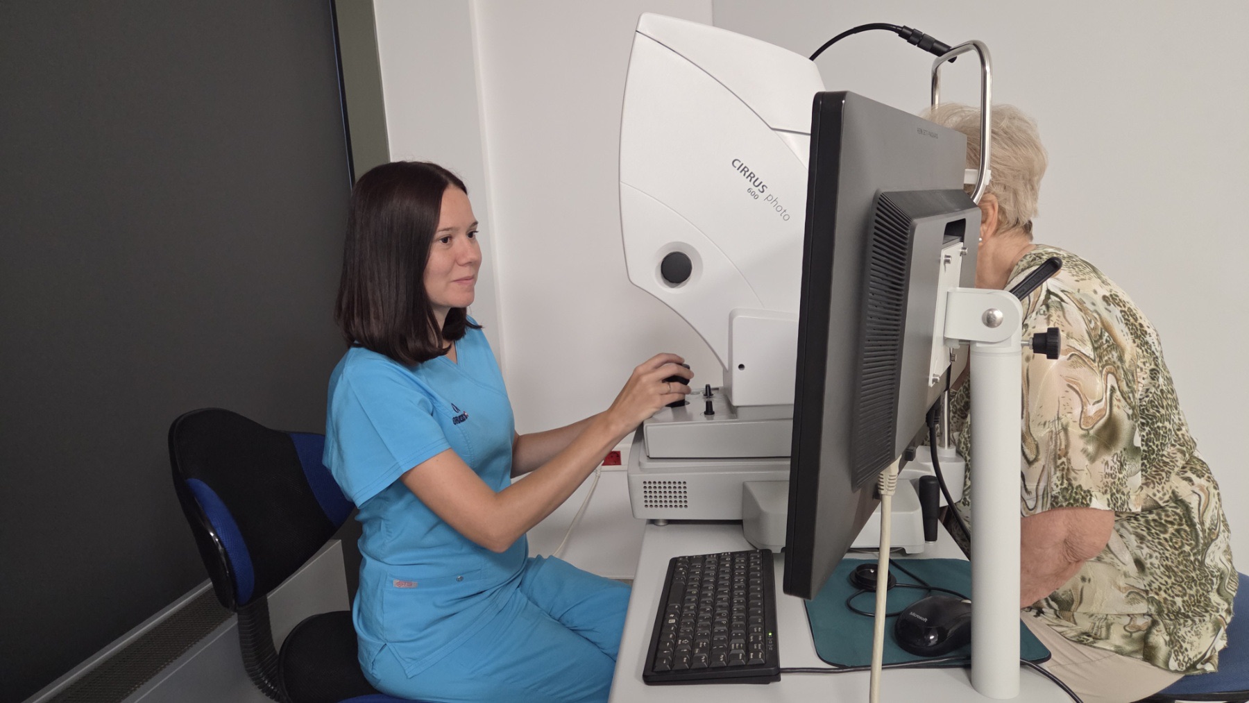
Optical coherence tomography is a non-invasive, non-contact, safe and rapid imaging method that produces high-resolution cross-sectional images of structures in the front and back of the eye. The OCT scanner is similar to a CT scanner in that it provides a detailed view of the tissues being examined. Its imaging resolution is considerably higher than that of ultrasound, which is why OCT is sometimes described as "in vivo histology".
OCT enables the diagnosis and monitoring of retinal complications caused by diabetes, retinal vascular occlusions sometimes referred to as an "eye stroke", age-related macular degeneration and many other conditions, including macular holes and epiretinal membranes. It is used to measure macular oedema precisely, monitor the response to treatment such as anti-VEGF therapy, and follow other changes in the macula.
OCT also supports the diagnosis and monitoring of glaucoma by measuring the retinal nerve fibre layer around the optic nerve. OCT angiography makes it possible to examine blood vessels without contrast dye. Imaging and analysis of the front of the eye with anterior segment OCT, or AS-OCT, is useful in diagnosing and managing corneal conditions such as keratoconus and corneal dystrophies. OCT is now an essential diagnostic tool in every modern ophthalmology clinic.
Appointments
Not sure which examination you need?
Tell us about your symptoms and our team will help you book the right first step.