
Eye clinic / patient information
Retinal detachment
The retina is the inner lining of the wall of the eyeball and is a neurosensory structure that responds to light stimuli. Light stimulation triggers a chain of chemical processes in the retina and creates electrical impulses that travel through the optic nerve and visual pathway to the brain.
The retina consists of several layers, traditionally described as 10 layers. These can be divided into the inner neurosensory part and the outer layer known as the retinal pigment epithelium (RPE).
What is retinal detachment?
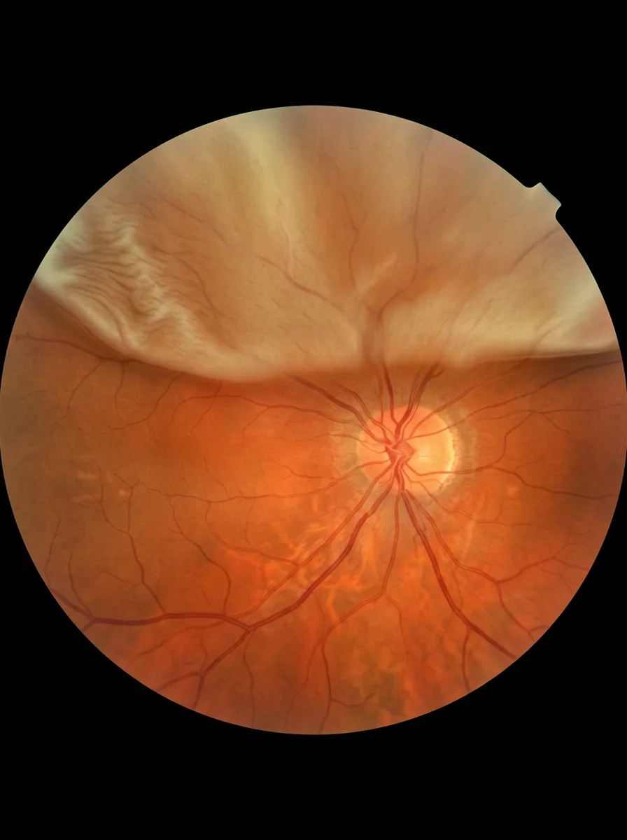
This pathological process is the separation of the neurosensory retina from the retinal pigment epithelium.
There are several types of retinal detachment, classified according to the mechanism by which they develop. A detachment caused by pre-existing retinal tears is called a rhegmatogenous retinal detachment. Detachments caused by other mechanisms are classified as non-rhegmatogenous retinal detachments.
Risk factors for retinal detachment
A number of factors may predispose a person to retinal detachment. These include short-sightedness, previous cataract or glaucoma surgery, previous eye injury, retinal detachment in the other eye, retinal detachment in parents or relatives, the use of eye drops in glaucoma treatment, and changes at the back of the eye associated with diabetes.
What are the symptoms, and when should you contact an ophthalmologist?
The appearance of flashes of light, floaters, a web, a curtain or tobacco-smoke-like changes in the visual field are signs that require an ophthalmic examination.
How is retinal detachment treated?
Retinal tears can be treated with laser photocoagulation before a retinal detachment develops.
Retinal detachment is treated only with a surgical procedure, such as pars plana vitrectomy or scleral buckling.
Appointments
Not sure which examination you need?
Tell us about your symptoms and our team will help you book the right first step.