
Diagnostics / preparation for surgery
Ocular biometry
Precise measurement of the length of the eye and of keratometry, which gives the doctor the basis for calculating the intraocular lens before cataract surgery.
What the doctor measures and why it matters
Ultrasound biometry measures the axial length of the eye. That value, together with other measurements of the anterior segment of the eye, is used to calculate the power of the intraocular lens implanted during cataract surgery.
The A-scan records the echo of the ultrasound wave at the boundaries of the ocular structures. The software of the device then uses the values obtained as part of a calculation that the doctor checks and interprets in the context of the whole examination.
What the measurement looks like
The examination is planned as part of the preparation for a precise choice of intraocular lens.
Preparing the eye
The medical team prepares the patient and explains the position during the measurement.
A-scan measurement
The probe is directed along the axis of the eye so that the device records the echoes of the ocular structures.
Checking the result
The doctor assesses the quality of the measurement and uses the data in planning the operation.
Two measurement techniques
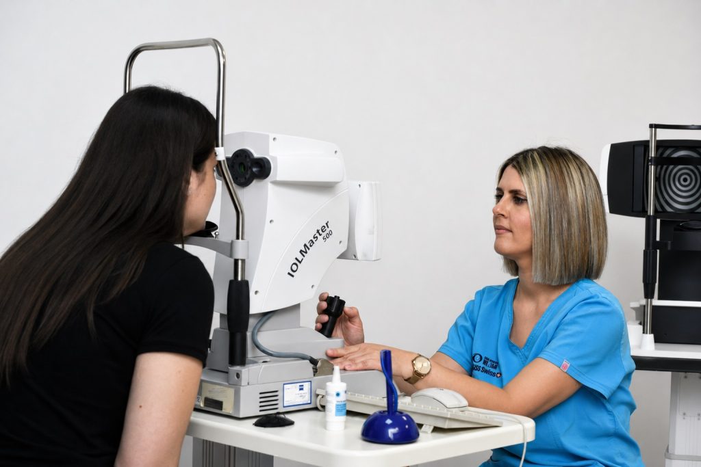
Optical biometry (IOL-Master) works on the principle of low coherence optical reflectometry. It is a non-contact scan of the eye in which a single click provides the values of the ocular parameters: keratometry, the position of the steepest meridian of the cornea, the depth of the anterior chamber and the axial length of the eyeball. Based on these parameters, and using the appropriate formulas, the software of the device determines the power of the intraocular lens (IOL) to be implanted during cataract surgery.
Ultrasound biometry is the measurement of the axial length of the eye (Lax) using ultrasound waves. A one dimensional A scan is used. There are 2 methods: immersion, in which the ultrasound probe is immersed in a water bath placed on the cornea, and contact, in which the probe is placed on the top of the cornea. By entering the keratometry values into the device, its software determines the power of the intraocular lens (IOL) to be implanted during cataract surgery.
Non-contact scan of the eye on the IOL-Master, a single click provides the ocular parameters.
Measurement of the axial length of the eye with an A scan probe, using the immersion or the contact technique.
The value only makes sense together with the other measurements and the ophthalmologist examination.
VIDAR-ORASIS SWISS / Novi Sad
Do you need biometry or preparation for surgery?
Talk to our team. You will be told which examination to book and what you need to bring.