

There is one conversation that repeats itself in the eye clinic in almost the same words. A patient comes in, sits down and says: "Doctor, I had surgery a year ago, I could see perfectly, I read without distance glasses, I drove at night without any problem. And now it has gone blurry again. Has my cataract come back? Has something gone wrong with the lens? Did I do something wrong?"
Posterior capsule opacification and YAG capsulotomy are something we see from time to time at the Vidar-Orasis Swiss Special Eye Hospital in Novi Sad, because it is a possible development after cataract surgery. The hospital has been working since 2010; the team includes, among others, four university professors. What follows is the explanation a patient receives at a check-up, only written down so that it can be read in peace.
That question usually carries more anxiety than the questions the patient asked before the surgery itself. Once you have been through the decision, through the waiting, through the day of the procedure and through the weeks of recovery, and once you have finally got a clear picture back, the thought that all of it is going back to the beginning is hard to take. Many people describe that feeling as disappointment rather than as a health problem.
That is why this text begins with the answer and not with an introduction. Cataract does not come back. The clouded natural lens that was removed during the surgery cannot grow again, and the artificial lens implanted in its place does not become cloudy over time. What does become cloudy in some patients is the thin transparent membrane in which that artificial lens sits, the so called posterior capsule. In everyday speech this is called secondary cataract, in reports it is often written as posterior capsule opacification, and it is resolved with a short laser procedure called YAG capsulotomy.
The name "secondary cataract" is historical and somewhat unfortunate, because it immediately suggests to the patient that the cataract is back. It is not. This is a change in a completely different structure, not a return of the disease. It is not the surgeon's mistake, it is not the patient's mistake and it is not a sign that the surgery was unsuccessful. It is a common and expected development that accompanies modern cataract surgery in a number of people, and one that patients unfortunately are often not told enough about before the surgery.
Below you will find what the capsule actually is and why it is deliberately left in the eye, why it becomes cloudy after some time, when this happens, how you will recognise that this is exactly what is going on, what YAG capsulotomy looks like, what happens after the procedure, which signs should send you to a doctor immediately and how much it costs. Everything written here is general information that does not replace an examination, because vision after cataract surgery can become blurred for several reasons, and which one is yours can be established only by an examination of the eye.
What actually happened in your eye during the surgery
For the story about secondary cataract to make sense, it helps to see what was left in the eye after the surgery.
The natural lens of the eye sits behind the pupil, that black round opening in the middle of the iris. In a young person it is transparent and elastic, and its job is to cast a sharp image onto the retina, the thin layer of nerve tissue on the back wall of the eye that does what the sensor does in a camera. A cataract is not a deposit over the eye and it is not a membrane growing across the pupil, even though many people imagine it that way. A cataract is a clouding of that lens itself: the proteins inside it change their structure, the lens loses its transparency and light passes through it scattered. If you are interested in what happens with vision before the surgery, the cataract page covers that in more detail.
Here comes the part that is key to understanding secondary cataract. The natural lens does not sit freely in the eye. It is housed in a very thin, transparent, elastic bag that wraps around it on all sides. That bag is called the lens capsule. Picture a large grape: what you eat is the contents, and what stays between your fingers when you peel the grape is the skin. The capsule is that skin, only incomparably thinner and completely transparent. It has a front wall, which faces the pupil, and a back wall, which faces the retina and which is extremely thin.
During modern cataract surgery the surgeon enters the eye through a very small incision at the edge of the cornea (the transparent front part of the eye, which you can picture as the glass on a wristwatch), makes a circular opening in the front wall of the capsule, breaks the clouded lens into tiny pieces with an ultrasound probe through that opening and removes them. That technique is called phacoemulsification and in the literature it is described as the standard method, which is a fact your doctor can confirm for you. What is deliberately not touched is the back wall of the capsule. It stays in place, whole, transparent and stretched like the skin on a drum.
An intraocular lens is then implanted into that emptied bag, an artificial lens that stays in the eye permanently. It is folded and slipped in through the same small incision, and inside it unfolds and rests against the capsule. The capsule holds it in the same plane in which the natural lens was. The cataract surgery page covers the procedure itself and the preparation for it in more detail.
Why the capsule is left in the eye at all
Patients who hear this explanation often ask a completely logical question: if that very membrane becomes cloudy later, why is it not removed straight away, during the surgery, and the problem solved in advance?
The answer is that the capsule is not a forgotten leftover but a load bearing part of the construction. It has at least three jobs.
First, it holds the artificial lens in place. For vision to be sharp, the lens has to sit at an exactly defined depth, centred on the pupil and without tilting. The capsular bag surrounds and stabilises it. Without it the lens would have to be fixed in some other way, and every such option is more complex and carries more risk.
Second, it separates the front and the back part of the eye. Behind the capsule is the vitreous body, a transparent gelatinous mass that fills most of the eyeball, and behind it the retina. An intact posterior capsule is the natural partition between those two zones. When that partition is missing or when it is damaged, the vitreous body can move forward, and that changes the relationships inside the eye and is linked with a higher risk of problems in the retina.
Third, it preserves the shape and the position of everything else. The bag is anchored by fine fibres to the ciliary body, a ring of muscle tissue around the lens, and that system keeps the whole optics of the eye in balance. On top of that, precisely because the contents of the bag are removed while the bag stays, the surgery can be short and can be done through a small incision, without stitches.
Leaving the posterior capsule is therefore not a compromise but the condition for the surgery to be what it is. The price of that solution is that in a number of people that same capsule loses its transparency over time, and that is resolved with a short laser procedure without entering the eye.
Why the posterior capsule becomes cloudy and why this is not a new cataract
Now we come to the phenomenon itself. When the contents of the lens are removed, a small number of the eye's own lens cells always remain on the inner side of the capsule. That is not an oversight by the surgeon. Those cells are microscopic, pressed against the wall of the bag, especially in the corner where the front and the back wall meet, and it is not possible to remove them down to the last one without damaging the capsule itself.
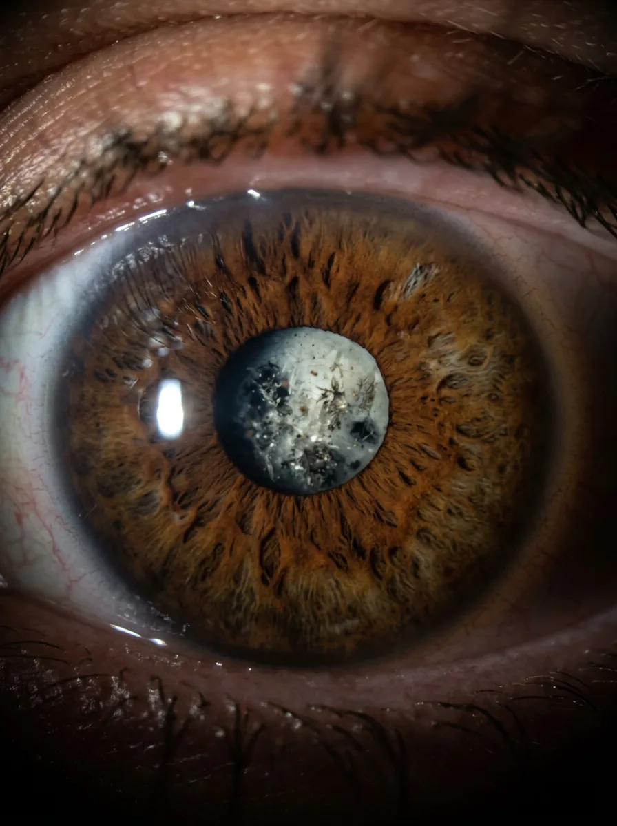
Those remaining cells are alive and they divide. In an empty bag with an artificial lens they carry on doing what lens cells normally do: they multiply and migrate across the inner surface of the capsule. In some people a part of those cells changes and begins to create connective fibres, so the capsule shrinks and wrinkles. The result is that the once transparent posterior capsule becomes cloudy, wrinkled or dotted with tiny bubbles.
The literature describes two basic appearances of that change: one looks like tiny transparent beads lined up along the membrane, the other is hazy and wrinkled. It is not unusual for both forms to be seen in the same eye. For the patient this division has no practical significance, because both types are resolved with the same procedure, but it explains why some people describe their complaints more as haze and others more as glare and doubling of the image.
The essential point worth repeating: this is not a cataract. A cataract is a clouding of the lens, and that lens is no longer in the eye. The artificial lens is made of a material that does not change its transparency over time. What is cloudy is the membrane behind it. The difference is roughly like the difference between a dirty pane of glass in a window and a dirty curtain behind that pane: the picture is blurred in both cases, but they are cleaned in completely different ways.
For the same reason secondary cataract is not a sign that the surgery was done badly. It is a biological response of the tissue, similar to the way the skin creates a scar at the site of a cut. It is not a matter of how careful you were, whether you lifted weights, read too much or looked at your phone. Nothing you did after the surgery caused it and nothing you might have done differently would have prevented it.
Who more often develops posterior capsule opacification
Not everyone develops it and it does not appear in everyone at the same time. How common it is exactly and within what period it most often appears is something your doctor should tell you, because the figures in the literature differ according to the generation of lens and the technique, and the documentation of the Vidar-Orasis Swiss clinic does not state a specific percentage. What is certain is that the phenomenon is common enough that every operated patient should know that it exists, and routinely enough resolvable that it should not be a source of fear.
The literature links the following circumstances with more frequent or earlier clouding of the capsule, and each of them should be understood as a tendency and not as a rule.
Younger age at the time of the surgery. The cells of a younger eye are more active and divide faster. That is why in children and young people operated on because of congenital or traumatic cataract the clouding of the capsule appears more often and earlier, so in them a different approach is sometimes taken during the surgery itself.
Accompanying diseases of the eye. Especially conditions that come with inflammation inside the eye, such as uveitis, then diabetes, as well as conditions that were changing the relationships inside the eye even before the surgery.
The course of the surgery itself. In a more complex procedure, for example with a very hard cataract or in an eye with weaker fibres that hold the capsule, the possibility that the capsule will react later is greater.
The type and the shape of the implanted lens. The material of the lens and the shape of its edge affect how easily the cells spread across the back wall of the capsule. Lenses with a sharp edge are linked in the literature with a lower frequency, because that edge creates a mechanical barrier.
None of the above is something the patient can influence after the surgery. There is no diet, drops, vitamin, exercise or way of life that prevents or slows the clouding of the capsule. If you come across a claim that any of this helps, treat it with caution.
When it appears
The timing is the biggest source of confusion, because secondary cataract almost never appears immediately.
The typical sequence looks like this. After the surgery vision improves and stabilises over a few weeks. The patient goes through the check-ups, gets new glasses if they are needed, and enters a period in which they see better than for years before. That period lasts for months, often for years. And then, gradually and imperceptibly, the picture begins to fade. Few people can say the exact date when it started.
Because of that gradualness many patients stay silent about it for a long time. Some think their glasses have got weaker, so they go to the optician. Some think they have grown older and that this is normal. Some put off the examination out of fear of hearing bad news. And some, and this is not rare, think that their surgery has "stopped working".
So it is worth putting it this way: clouding that appears months or years after the surgery, gradually, without pain and without redness, most often turns out to be posterior capsule opacification. That is resolved with one short outpatient procedure, so there is no reason to live with it or to put off the examination.
The opposite situation should be clearly separated. If vision worsens suddenly, within one day or a few hours, with pain, strong redness, a shadow coming in from the side or a burst of flashes and floaters, that is not the picture of secondary cataract and it does not wait.
Symptoms: how you will recognise it
The complaints in posterior capsule opacification resemble the complaints you had before cataract surgery, and that is one of the reasons why people conclude that their cataract has come back. Here is how it looks in everyday life.
Gradual haze. The picture is as if through fogged glass: it is not dark, but hazy. Contrasts weaken, so it is harder to make out the edge of a step or a kerb.
Glare and reflections, especially at night. Often the first symptom people notice. Headlights from the opposite direction make star shaped trails and halos, street lighting spreads out, and driving at night becomes unpleasant.
Paler colours. White starts to look like dirty white or greyish, and the difference between dark blue and black becomes hard to notice. Many people notice this only when they close one eye and compare.
Harder reading, especially in weaker light. The letters stay the same size but look faintly printed. You need stronger light than before for the same page or for the leaflet with a medicine.
The impression that your glasses are constantly dirty. The classic situation: a person wipes their glasses several times a day, changes the cloth, buys a cleaning fluid, and only then starts to suspect that the problem is not in the lens.
A double or smeared image in one eye. When you close the other eye, the outline of letters or headlights has a shadow beside it. That happens because the wrinkled capsule scatters light.
A difference between the two eyes. If the capsule has clouded in only one eye, the difference is noticed by closing the eyes in turn. Many people come for an examination precisely because they happened to cover one eye and were frightened by how much worse the picture in the other one was. At the examination this is seen as a drop in the number of lines you can read on the chart.
What is absent in secondary cataract matters just as much: there is no pain, no redness, no discharge, no swelling of the eyelid. If any of that is present, it is something else and you should seek help immediately.
What else can blur vision after cataract surgery
Blurred vision after cataract surgery does not have one cause but a whole group of possible reasons. Some are harmless, some require treatment, and some require an urgent reaction. Only an examination can tell them apart.
Posterior capsule opacification. Gradual, without pain, months or years after the surgery. Resolved with a laser.
A change of prescription and the need for new glasses. It is not rare that after some time a correction for distance or for near is needed, or that the existing glasses no longer fit.
Dry eye. A very common and underestimated cause. Vision blurs from time to time, improves after blinking and is worse towards the end of the day. Blurring that comes and goes in waves points more to the tear film than to the capsule.
Swelling of the centre of the retina. A build-up of fluid in the macula, the part of the retina responsible for sharp central vision. It gives blurring and distortion of straight lines and requires treatment.
Diseases of the retina and the macula. Age related macular degeneration and diabetic changes exist independently of the cataract and can progress after the surgery. That is also the reason why a particular sharpness of vision is never promised before the surgery: the final result depends on the state of the other parts of the eye.
Glaucoma, changes on the cornea and displacement of the implanted lens. Damage to the optic nerve gives no symptoms for a long time, swelling of the cornea also gives haze, and displacement of the lens is rare but possible, especially after an injury to the eye.
Retinal detachment. An emergency. It gives a shadow that enters the field of vision, with flashes and a burst of floaters.
That is why an examination is done before every laser procedure. If the cause is not the capsule, the laser will not help; and if the capsule is treated without insight into the state of the retina, the real reason for poor vision is missed.
How the diagnosis is made
The examination with this suspicion is a usual ophthalmological examination, with a few points of emphasis.
The conversation. The doctor wants to know when you were operated on, how you saw after the surgery, when the clouding started, whether it is getting worse gradually or suddenly, whether it is in one eye or both, and whether you have pain, redness, flashes or a shadow in the field of vision. It is useful to think through the sequence in time before you go.
Checking the sharpness of vision and measuring eye pressure. Vision is checked with glasses and without them, so that it can be seen how much the situation has changed compared with the findings after the surgery. That is why it is good to bring your old documentation.
Examination on the slit lamp. Through a device with strong illumination and magnification the doctor looks at the front part of the eye and the position of the artificial lens. A clouded posterior capsule is seen as a hazy or wrinkled membrane immediately behind the lens.
Examination of the fundus with a dilated pupil. Drops that widen the pupil are instilled, and after some twenty minutes the retina is examined. This part is compulsory, because it checks whether behind the capsule there is another reason for poor vision.
Additional tests if needed. Most often imaging of the retina, if the doctor wants to rule out swelling of the centre of the retina or other changes. In the price list of the Vidar-Orasis Swiss clinic such imaging exists as a separate service.
After the pupil is widened vision is blurred and sensitive to light for a few hours, so it is not recommended that you drive yourself from the examination. Plan for company or for transport.
Book an examination by calling 021 63 61 222. If your vision has gradually become blurred after an earlier cataract surgery, an examination is the only way to establish whether the reason is a clouded capsule or something else. Vidar-Orasis Swiss works on weekdays until 8 pm and does not work at weekends.
YAG capsulotomy: what it looks like
When it is established that the cause is a clouded posterior capsule and that there is no other reason for poor vision, the solution is a procedure called YAG capsulotomy. "YAG" is an abbreviation for the type of crystal in the laser (yttrium aluminium garnet), and "capsulotomy" means making an opening in the capsule.
The most important thing is to understand that this is not surgery in the classic sense. There is no incision, no instrument enters the eye, there are no stitches, you do not go into an operating theatre and you do not stay in hospital. The laser beam passes through the cornea and through the artificial lens, and delivers its energy exactly at the depth of the clouded capsule.
The principle is simple. Instead of the clouded membrane (the capsule) being cleaned or removed, an opening is made in it in the middle, exactly behind the pupil, in the zone through which the light that makes the image passes. The clouded part stays to the side, outside the optical axis, where it does not interfere, and the edges of the capsule still hold the artificial lens in place.
Preparation
The preparation is short. It is usual for drops that widen the pupil to be instilled before the procedure, because through a wide pupil the whole clouded membrane (capsule) is better seen and it is easier to aim. The drops start to work within about fifteen minutes. Whether the procedure at the Vidar-Orasis Swiss clinic is done on a widened pupil is something your doctor will confirm for you.
Company is planned for the same reason. After the pupil is widened vision is blurred and the eye is sensitive to light for a few hours, so driving in that state is not advisable. The simplest thing is for someone to bring you and take you home. Bring sunglasses as well.
It is also usual for a local anaesthetic to be instilled, because a special contact lens is rested on the eye which helps the laser beam to be aimed precisely.
The procedure itself
You sit in front of a device that from the outside resembles the instrument the doctor usually examines you with. You rest your chin on the support and your forehead on the band, and you will be told to look at one point and to keep still.
Then follows a series of short laser pulses. They are heard as a quiet crackling, and each one makes a microscopic opening in the membrane (capsule); by lining up those points a circular opening is made. Most people see flashes of light and a red aiming dot while this is going on. For most the sensation is bearable and is described as slight pressure of the contact lens together with discomfort from the bright light, while in a smaller number of people a brief stinging occurs.
The procedure itself is short, in the literature it is described as a procedure of only a few minutes per eye, and most of the time is taken up by the preparation and by waiting for the drops to work.
If you move or blink in the middle of the procedure, that is not a problem. The device works in pulses, so the doctor simply waits for you to settle. If a sufficiently large opening is not achieved, the procedure can be completed at another appointment.
A few things patients expect and which do not happen: you do not come on an empty stomach unless the doctor says otherwise, therapy for blood pressure, heart or sugar is not interrupted without agreement with the doctor, no incision is made, no bandage is put over the eye and you do not stay overnight for observation.
After the procedure
The first hours
Immediately after the procedure vision is most often blurred, for two reasons: the pupil is wide from the drops, and tiny particles created while making the opening are floating in the eye. Both pass.
Eye pressure rises temporarily after YAG capsulotomy in some patients, so it is usual for it to be measured immediately after the procedure or during the same day. Whether pressure lowering drops are given preventively at the Vidar-Orasis Swiss clinic is something your doctor should tell you.
In most patients vision clears during the same day or over the next few days, as the pupil narrows and as the particles in the eye settle. How quickly this happens in an individual depends on the state of the eye and on how dense the membrane was, so that too is a question for the doctor. A particular sharpness of vision is not promised, because the final result depends on the state of the retina, the optic nerve and the cornea. If there was a disease of the macula or the optic nerve before the procedure, the opening in the capsule removes only one obstacle, not the others.
The first days and weeks
It is usual for drops to be used for some time after the procedure. Anti-inflammatory drops are most often mentioned, and sometimes drops for pressure as well, but the exact schedule and duration are determined by the doctor and this differs from patient to patient. Do not stop the drops on your own as soon as you feel better and do not extend them beyond the recommended time.
The restrictions are, compared with cataract surgery, much smaller, precisely because there is no incision, so you usually return to everyday activities the same or the next day. Ask your doctor whether and for how long you should avoid physical effort, bending over, the swimming pool or sport.
A common and unsettling development after the procedure is new floaters in the field of vision: tiny dots, threads or specks that float and move with your gaze. They arise from particles of the clouded membrane that drift into the vitreous body, and in most people they become less noticeable over time. That is a usual consequence of the procedure, but there is a limit that must not be overlooked: a sudden burst of many new floaters, especially together with flashes of light or a shadow at the side, is not usual and calls for an urgent examination. The text on floaters and flashes speaks about the difference between harmless and worrying floaters, and it is worth reading before the procedure, so that you know what you are watching for.
A follow-up examination is usually scheduled within a few weeks, and in people with glaucoma or other accompanying conditions it may be earlier and more frequent. Your doctor will tell you the schedule of check-ups that Vidar-Orasis Swiss applies.
When a new prescription is measured
If a clouded capsule has weakened your vision for distance or for near, wait for the situation to settle before you order new glasses. Measuring a prescription while the pupil is wide and while vision is still clearing gives an unreliable result. Your doctor will tell you when the right moment is.
Warning signs after the procedure
These signs do not belong to the usual course after YAG capsulotomy. If any of them appears, do not wait for the scheduled check-up and do not assume that it will pass by tomorrow.
- A sudden drop in vision. Not gradual clearing with ups and downs, but a marked and sudden worsening.
- Flashes of light ("lightning"), especially in the periphery of the field of vision, in the dark or on a sudden movement of the head.
- A sudden burst of new floaters, considerably more than there were immediately after the procedure, or a burst that looks like a shower of soot or a swarm.
- A shadow or "curtain" that enters the field of vision from the side, from above or from below, or a part of the picture that is missing.
- Severe pain in the eye that does not let up, especially together with nausea, vomiting and headache.
- Marked redness of the eye with discharge, swelling of the eyelid, stuck together lashes.
- Distortion of straight lines: a door frame, the edge of a tile or a line on paper looks wavy or broken.
If anything from this list appears, call 021 63 61 222. Vidar-Orasis Swiss works on weekdays until 8 pm and does not work at weekends. Outside those hours contact the nearest emergency service or an on-call eye clinic. When these signs are present it is better to come unnecessarily than to be too late.
Possible complications and why the procedure is not done "just in case"
YAG capsulotomy is performed very often in practice and is considered a routine procedure. But no procedure on the eye is without possible unwanted outcomes, and it is fair that you should know them before you agree to it.
A temporary rise in eye pressure. The most common occurrence, usually short lived, which is why pressure is measured after the procedure. In people with glaucoma it is monitored more carefully.
An inflammatory reaction in the front part of the eye. Usually mild and passing, which is why anti-inflammatory drops are given.
Floaters in the vitreous body. Usual, and they generally weaken over time.
Swelling of the centre of the retina. A build-up of fluid in the macula, which gives blurring and distortion of the image and requires treatment.
Damage to or displacement of the artificial lens. Rare. A tiny mark from a laser pulse can remain on the lens, which most often does not affect vision.
Retinal detachment. A rare but serious possibility, and the main reason why after the procedure recognising flashes, floaters and a shadow is insisted on.
Because of all this the procedure is not done just because the capsule is not perfectly transparent at the examination. It is done when the clouding really interferes in everyday life or when it prevents the doctor from monitoring the retina, for example in a patient with diabetes. If you see well and the capsule is only slightly hazy, it is reasonable for that to be followed at check-ups. The same principle applies to the decision about cataract surgery itself, which the text about when a cataract is operated on speaks about.
The decision is a joint one. You describe how much it bothers you, the doctor assesses the findings and the risks in your particular case.
Is the procedure repeated
The question is logical: if the membrane clouded once, will it cloud again?
The essence is that in this procedure the clouding is not removed from the membrane, but an opening is made in it. The opening is a physical hole and it stays. That is why, when it has been made in a sufficient size and in the right place, the same zone is not expected to become cloudy again.
There are nevertheless situations in which the patient comes back for a top-up: if a smaller opening was made the first time so that when the pupil widens in the dark its edge falls within the field of vision, if the edges wrinkle over time, or if in particularly prone eyes the cells spread along the edges as well. Whether and how often the procedure is repeated in practice is something your doctor should tell you.
The following is also important: if vision becomes blurred again several years after a successful capsulotomy, the first thought should not be that the membrane has come back, but that there is another cause. A repeat examination is the only way to tell those apart.
Price
In the price list of the Vidar-Orasis Swiss clinic YAG capsulotomy is listed at a price of 18,000 RSD. The same price list also contains Nd:YAG iridotomy at a price of 18,000 RSD, which is a different procedure with the same laser and the two should not be mixed up.
Two notes go with that price. First, the price of the procedure does not include the examination that establishes that the clouded capsule really is the cause of the complaints; the ophthalmological examination is listed separately in the price list, as is additional imaging. If you want to know in advance how much the whole visit will cost, ask about that when booking and say that you are coming because of blurred vision after an earlier cataract surgery.
Second, the price list has a date of the last change and prices are subject to change, so always check the current price by calling. You can see an overview of current services on the services and prices page. Whether the procedure is charged per eye, whether there is a difference in price for a top-up of an opening made earlier and whether the follow-up examination is charged separately are questions for the clinic when booking.
How it looks in everyday life
A description of symptoms often misses what people actually recognise. Here are the situations in which clouding of the capsule is most often noticed.
Driving at night. The situation in which most people take fright for the first time. Headlights from the opposite direction make stars and halos, wet asphalt glitters, the markings on the road are harder to see. Many people stop driving at night because of this and do not tell anyone why. That is not a matter of age and it is not something you should accept until it has been checked. The same goes for driving in the afternoon towards a low sun, when the picture turns into a white haze.
Reading a label in a shop. Small print on a box of medicine is a classic test. The lighting in supermarkets is strong but diffuse, and that is exactly what creates the problem with scattering of light.
Working at a computer. The screen becomes hazier towards the end of the day, letters seem as if they have a shadow. Here it is important to separate clouding from the capsule, which is constant, from dry eye, which improves temporarily after blinking.
Recognising faces. You recognise an acquaintance in the street with more difficulty from the distance at which you used to recognise them, and the expression on the face of the person you are talking to looks "unclear".
Television, fine handwork and work in the garden. The ball gets lost in the broadcast, subtitles are read with more difficulty, threading a needle becomes a struggle, and in the garden it is harder to tell a young plant from a weed.
Looking after grandchildren. Following a child on the playground, reading the small letters in the book the child brings, pouring medicine into a spoon.
Stairs and kerbs. Weaker contrast means that the edge of a step is harder to make out, which increases the risk of a fall. This is the reason why blurred vision in older people is not viewed only as a question of comfort.
If you recognise yourself in several places on this list, that is reason enough to book an examination, regardless of how well you saw immediately after the surgery.
What to ask your doctor
It is good to come to the examination with a list, because in the consulting room questions are easily forgotten.
- Is the cause of my blurring really the posterior capsule, or is there another reason as well?
- What is the state of my retina and optic nerve?
- Do I need the procedure now or can it wait and be monitored?
- How much improvement in vision can I realistically expect in my case?
- Is the procedure done on a widened pupil and do I need someone to come with me?
- How long after the procedure should I not drive?
- Which drops do I get after the procedure, for how long and on what schedule?
- When is the check-up and what is checked at it?
- Which signs mean that I have to seek help immediately?
- Do my other illnesses or medicines change the plan?
- How much does the procedure cost, and how much the examination and the imaging?
- When can I have my prescription measured for new glasses, and is the procedure done on both eyes on the same day?
Bring your old documentation as well: the discharge letter, the details of the implanted lens, the findings of check-ups and a list of the medicines you take.
Book an examination on 021 63 61 222. If your vision has gradually become blurred after an earlier cataract surgery, an examination establishes whether the cause is a clouded posterior capsule and whether the procedure is justified in your case. Vidar-Orasis Swiss works on weekdays until 8 pm and does not work at weekends.
Frequently asked questions
Has my cataract come back?
No. The clouded natural lens has been removed and cannot grow again, and the implanted artificial lens does not develop a cataract. What becomes cloudy is the thin membrane on which that lens sits. The name "secondary cataract" is historical and confusing, but it does not describe a return of the same disease.
Is it a sign that the surgery was done badly?
It is not. The posterior capsule is deliberately left because it holds the artificial lens and separates the front and the back part of the eye. The clouding arises as a biological reaction of the remaining cells and does not depend on how carefully the procedure was performed.
How long after the surgery does it usually appear?
Most often not immediately, but months to years after the surgery, and gradually. Precise figures on frequency and on the time of appearance should be given to you by your doctor.
Can it be treated with drops or exercises?
No. There are no drops, supplements, diet or exercises that remove the clouding of the capsule. The only solution is making an opening with a laser.
Does YAG capsulotomy hurt?
The procedure is done under drops, with a local anaesthetic that numbs the surface of the eye. Most patients describe a feeling of pressure from the contact lens on the eye and discomfort from the bright light. In a smaller number of people a brief stinging or a mild headache occurs. If you have a low threshold of tolerance for discomfort, tell the doctor in advance.
When will I see better?
In most patients vision clears during the same day or over the next few days, as the pupil narrows and as the tiny particles in the eye settle. How sharp your vision will be also depends on the state of the retina, the optic nerve and the cornea, so the final outcome is not promised in advance.
May I drive home myself?
It is not recommended. The pupil stays wide for a few hours, vision is blurred and the eye is sensitive to light. Plan for company or for transport and bring sunglasses.
Are drops given after the procedure?
It is usual for anti-inflammatory drops to be used for some time, and in some patients drops for eye pressure as well. The exact schedule and duration are determined by the doctor; do not stop them on your own.
Does the procedure have to be repeated?
The opening made in the membrane (capsule) is permanent and the same zone is not expected to become cloudy again. In a smaller number of cases a top-up is done, for example if the opening was too small or if the edges wrinkle.
Can I postpone the procedure?
If the clouding does not significantly bother you in everyday life and if the doctor can monitor the state of the retina, monitoring is a reasonable choice. The procedure is done when the complaints really interfere or when they prevent the doctor from examining the fundus.
I see floaters after the procedure, is that normal?
Tiny dots and threads are usual after capsulotomy and in most people they become less noticeable over time. But a sudden burst of many new floaters, especially together with flashes of light or a shadow at the side, is not usual and calls for an urgent examination.
Is this the same as cataract surgery?
It is not. Cataract surgery is a surgical procedure with an incision, removal of the lens and implantation of an artificial one. YAG capsulotomy is a short laser procedure without an incision. The recovery is significantly different too; what recovery after the cataract surgery itself looks like is described in the text on recovery after cataract surgery.
What if I have glaucoma or diabetes?
Tell the doctor before the procedure. With glaucoma eye pressure is monitored more carefully after the procedure. With diabetes the state of the retina also matters, because a clouded capsule can make examining it more difficult.
Note
This text is informative in character and does not replace an examination by an ophthalmologist. Blurred vision after cataract surgery can have several different causes, and which one is yours can be established only by an examination. The time frames, drop schedules and recommendations given are general guidelines; the instructions you receive from your own doctor take precedence over any text. Do not change and do not stop prescribed therapy on your own. The prices given in the text are subject to change and should be checked by calling.
Sources and further reading
The medical content was reviewed by Prof. Dr Mirko Resan. This information does not replace an eye examination or individual advice from an ophthalmologist.