
Eye clinic / patient information
Keratoconus
Information about the examination, diagnostics and the possible next steps at the VIDAR-ORASIS SWISS clinic.
Introduction
The cornea is a transparent, avascular (without blood vessels), multilayered structure of the front part of the eye responsible for the refraction of light rays. It has approximately the shape of a segment of a sphere, with a smaller thickness in the centre than at the periphery.
What is keratoconus?
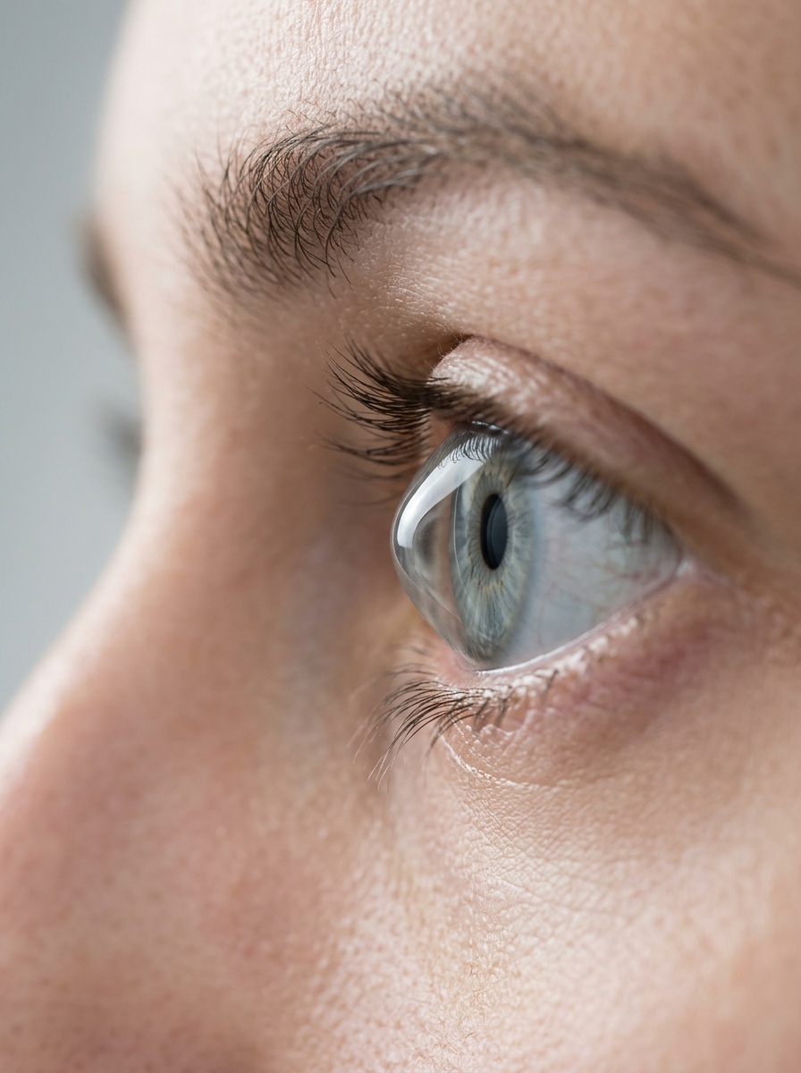
Keratoconus is a bilateral disease (ectatic dystrophy) which is characterised by a progressive reduction in the thickness of the corneal tissue which is accompanied by conical (cone shaped) bulging of the cornea with the greatest thinning at the apex of the cone. It develops as a result of genetic factors, enzymatic and chromosomal abnormalities, as a consequence of one of the surgical interventions, and other mechanical factors (frequent rubbing of the eyes). The rate of keratoconus in the general population is from 50-230 per 100,000 people, equally in both sexes.
At what age does it most often appear?
Keratoconus most often appears in the period of puberty, and it can manifest with unequal intensity in the two eyes. In the period between the ages of 30-40 there is a “slowing down” of the pace of the disease. This disease can be associated with other eye conditions such as congenital cataract, aniridia (poor development or absence of the iris), retinopathy of prematurity, retinitis pigmentosa, etc., as well as with systemic conditions (asthma) and various genetic disorders.
What are the symptoms and what is the prognosis of the disease?
The person notices a drop in visual acuity, an unclear image of perceived objects, increased sensitivity to light, the appearance of “double” images of perceived objects, increased watering of the eye.
If the progression of the disease is not stopped, there is a breakdown of the mechanisms that make the cornea resistant to water, which leads to excessive accumulation of water in the cornea, together with scarring, which endangers visual function.
Diagnosis
The ophthalmologist establishes the diagnosis on the basis of:
an examination (assessment of visual acuity, examination of all eye structures on the biomicroscope)
keratometry- a procedure where with a device we determine the curvature of the cornea
corneal topography-we measure the shape and size of irregularities in the curvature of the cornea, as well as its thickness (Orbscan)
OCT diagnostics of the anterior segment of the eye (AS-OCT)
Classification
On the basis of keratometric measurements and the shape of the bulging, we grade the severity of the disease from mild, moderate, marked, to very serious.
Treatment of keratoconus
The ophthalmologist carries out treatment according to the seriousness of the condition and the possibility of achieving the best correction of vision. At the beginning, if it is possible, we correct vision with glasses. A better solution for higher quality vision is correction with various types of contact lenses, with special methods of fitting them.
If keratoconus progresses and threatens greater thinning of the cornea, then it is necessary to carry out a surgical intervention by which the cornea will be strengthened („cemented“) and its further thinning prevented. That intervention is called Corneal cross-linking (CXL) and is a surgical method that we use when the disease is in the phase of progression and when a certain thickness of tissue still exists for this procedure. By the action of a vitamin B2 solution and a special UV lamp, we create “crosslinks” in the basic network of collagen fibres of the stroma, and thereby we increase the biomechanical stability of the cornea.
When the cornea is altered by scarring, then we proceed to an operative procedure which is called keratoplasty. We carry out removal of the diseased corneal tissue and replacement of it with full thickness donor tissue (penetrating keratoplasty). Recently the approach of lamellar keratoplasty (DALK) has also been used in specially selected cases.
Appointments
Not sure which examination you need?
Tell us about your symptoms and our team will help you book the right first step.